
Non-Surgical Joint Interventions: How Intra-Articular Injections Restore Synovial Fluid Function
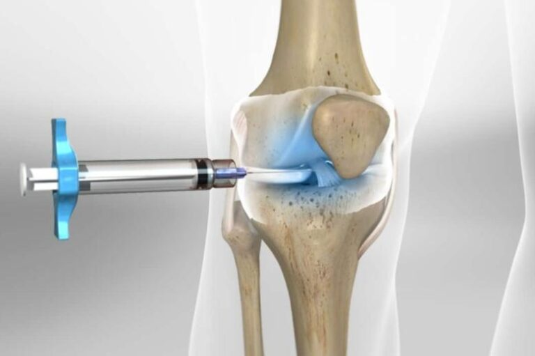
Symptomatic osteoarthritis of the knee routinely forces orthopedic clinics into a challenging management loop. Physical therapy builds supportive musculature, yet mechanical friction inside an inflamed joint continues to erode remaining hyaline cartilage. Oral analgesics temper localized discomfort. Unfortunately, long-term nonsteroidal anti-inflammatory drug (NSAID) use carries well-documented gastrointestinal, renal, and cardiovascular risks that often limit daily dosages in aging patient populations. When conservative oral regimens plateau and surgical joint replacement remains premature, intra-articular interventions serve as a intermediate biological treatment strategy.
To understand why synthetic viscosupplementation works, one must look closely at normal joint mechanics.
+———————————————————————————–+
| MECHANISMS OF VISCOSUPPLEMENTATION |
+———————————————————————————–+
| 1. Rheological Cushioning –> Restores temporary viscosity and elasticity |
| 2. Cellular Stimulation –> Triggers native synoviocytes to produce HA |
| 3. Receptor Binding (CD44) –> Downregulates inflammatory cytokine production |
| 4. Nociceptive Shielding –> Decreases mechanical stretch receptor firing |
+———————————————————————————–+
The Physiology of Synovial Fluid Degradation
Healthy synovial fluid relies on high concentrations of endogenous hyaluronic acid, a non-sulfated glycosaminoglycan synthesized continuously by specialized type B synoviocytes. These molecular chains reach massive weights between 4 and 10 million Daltons. Under low shear stress, such as static standing, these long-chain polymers entangle to form a thick, protective lubricant that prevents direct tissue contact. Under high shear stress, like running, the molecules alter their physical orientation to absorb structural impact.
Degeneration breaks this equilibrium.
In an osteoarthritic knee, elevated levels of inflammatory cytokines, including interleukin-1 beta and tumor necrosis factor-alpha, upregulate catabolic enzymes that chop native hyaluronan into fragmented, low-molecular-weight pieces. Concurrently, increased vascular permeability inside the synovial membrane dilutes the joint cavity. The concentration of functional hyaluronan drops by up to 67 percent. Deprived of its viscoelastic property, synovial fluid thins into a watery liquid that can no longer shield cartilage from mechanical friction.
How rapidly does this breakdown happen? Rapidly.
Once mechanical degradation outpaces biological synthesis, the joint enters a destructive feedback loop where physical wear triggers additional inflammatory signaling. According to clinical consensus statements published by the American Academy of Orthopaedic Surgeons (AAOS), loss of fluid viscosity correlates directly with increased chondrocyte apoptosis and progressive loss of joint space width visible on weight-bearing radiographs.
Mechanisms of Viscosupplementation and Clinical Sourcing
Intra-articular viscosupplementation counters this breakdown by injecting exogenous hyaluronic acid directly into the joint space. The primary objective involves restoring immediate rheological cushioning, but the physiological impact extends far past basic fluid mechanics.
Reintroduced hyaluronan binds directly to CD44 transmembrane receptors expressed on synoviocytes and chondrocytes. Binding inhibits the nuclear factor kappa B (NF-κB) signaling pathway, effectively downregulating matrix metalloproteinase production. Crucially, exogenous hyaluronan stimulates native synoviocytes to resume production of high-molecular-weight hyaluronic acid—or more precisely, it interrupts the local inflammatory signals that suppress endogenous synthesis. This biological signaling effect explains why clinical improvements often persist long after the injected compound has cleared the joint space through lymphatic drainage.
Selecting an appropriate hyaluronic acid formulation requires balancing molecular weight, cross-linking technology, and administration schedules. Standard preparations utilize either avian-derived hyaluronan or bacterial fermentation products. High-molecular-weight and cross-linked variations slow intra-articular degradation, reducing required clinic visits from five weekly sessions down to three or even a single injection.
In busy orthopedic practices, securing reliable inventory for these specialized biologic devices frequently creates administrative friction. Insurance pre-authorization requirements, fluctuating distributor stock, and cold-chain shipping logistics regularly delay patient care. When practice managers establish purchasing pipelines for specialized viscosupplements, reviewing verified medical supply channels ensures clinic schedules remain uninterrupted. Practice administrators evaluating inventory options can review professional Euflexxa ordering options to examine volume pricing, batch availability, and delivery terms directly. Maintaining consistent access to high-molecular-weight, non-cross-linked sodium hyaluronate formulations enables clinical teams to execute standardized, multi-dose injection protocols without running into supply chain bottlenecks.
Following successful procurement, precise anatomical placement determines clinical success.
Diagnostic Nuance and Patient Selection Frameworks
Not every patient complaining of deep joint pain benefits from intra-articular hyaluronan. Clinical efficacy depends heavily on structural selection criteria, baseline inflammation levels, and realistic therapeutic expectations.
|
Baseline Parameter |
Favorable Candidate Criteria |
Unfavorable Candidate Criteria |
Clinical Rationale |
|
Radiographic Severity |
Kellgren-Lawrence Grade II to III |
Kellgren-Lawrence Grade IV |
Requires preserved joint space; ineffective in complete bone-on-bone contact |
|
Inflammatory Effusion |
Minimal to mild joint effusion |
Severe, acute joint effusion |
Excessive fluid dilutes exogenous hyaluronan and elevates catabolic enzyme activity |
|
Previous Interventions |
Inadequate response to NSAIDs/PT |
Uncontrolled systemic disease |
Identifies failure of primary non-invasive management prior to viscosupplementation |
|
Primary Goal |
Function preservation & pain reduction |
Reversal of cartilage damage |
Sets realistic expectations; hyaluronan does not regenerate lost articular cartilage |
Is viscosupplementation overused in late-stage disease? Absolutely.
Many clinicians offer viscosupplementation as a compassionate attempt to postpone total knee arthroplasty in patients with complete loss of joint space. This practice wastefully inflates healthcare costs. Research synthesis by the Rheumatology Osteoarthritis Research Society shows that injecting hyaluronan into a bone-on-bone joint yields outcomes barely distinguishable from placebo injections. The mechanical cushion simply collapses under direct structural loading when no residual hyaline cartilage remains to absorb shear stress.
Texture of Clinical Practice: Injection Realities and Patient Misconceptions
Walk into any outpatient orthopedic clinic on a Tuesday morning, and you will hear the exact same set of questions from anxious patients. People consistently ask whether the needle will hurt, whether the fluid will rebuild their worn-out knee cartilage, and whether they can play tennis the following afternoon.
Setting clear physical expectations upfront prevents patient dissatisfaction down the road.
"Patients routinely assume hyaluronic acid works like joint spackle, physically filling in holes in their cartilage," notes an orthopedic clinic coordinator managing high-volume injection schedules. "We spend half our consultation time explaining that this treatment is a biological lubricant and anti-inflammatory signal, not a structural patch that reverses fifteen years of wear."
INJECTION WORKFLOW & PROTOCOL
+——————–+ +——————–+ +——————–+
| Pre-Infiltration | —> | Ultrasound-Guided | —> | Post-Procedure |
| Arthrocentesis | | Infiltration | | Restrictions |
| (Effusion removal) | | (Superolateral) | | (48hr light load) |
+——————–+ +——————–+ +——————–+
The physical injection protocol itself requires meticulous aseptic execution. While blind palpation-guided intra-articular injections remain common in primary care settings, ultrasound-guided infiltration significantly improves accuracy. Placement failure rates for non-guided knee injections reach up to 20 percent in patients with high body mass index or significant joint deformities.
When a joint presents with significant effusion, the clinician must perform a thorough arthrocentesis to aspirate excess synovial fluid before injecting the therapeutic compound. Skipping this aspiration step dilutes the exogenous hyaluronan immediately, rendering the treatment far less effective.
Following administration, patients must adhere to a strict 48-hour activity modification period. High-impact weight-bearing exercises, heavy lifting, and prolonged standing should be completely avoided. This brief resting period minimizes the risk of post-injection flare-ups, which present as acute joint pain, localized heat, and mild swelling within 24 hours of the procedure.
Comparative Evaluation: Corticosteroids vs. Hyaluronic Acid vs. Biologics
Clinicians managing osteoarthritic joints must choose between several distinct intra-articular modalities. Each therapeutic class features unique pharmacodynamics, onset speeds, and duration profiles.
Triamcinolone acetonide and other intra-articular corticosteroids provide rapid, potent pain reduction by shutting down localized inflammatory cascades. However, this relief is temporary, typically declining significantly after four to six weeks. Repeated corticosteroid administration poses serious risks to long-term joint health; high-frequency steroid injections accelerate chondrocyte toxicity and speed up articular cartilage loss, according to longitudinal imaging studies supported by the National Institutes of Health (NIH).
Platelet-rich plasma (PRP) represents an alternative biologic approach that utilizes concentrated autologous growth factors to stimulate tissue healing. While meta-analyses published by the Arthroscopy Association of North America (AANA) suggest PRP may yield longer-lasting pain relief than un-crosslinked hyaluronic acid in early-stage disease, standardizing PRP remains a regulatory nightmare. Platelet yield, leukocyte concentration, and activation protocols vary wildly between commercial centrifugation systems, making consistent clinical outcomes difficult to replicate across different practice locations.
Viscosupplementation occupies a balanced middle ground between these options.
Unlike corticosteroids, high-molecular-weight hyaluronic acid exhibits a favorable safety profile upon repeated administration, showing zero chondrotoxic effects over multi-year treatment cycles. While its onset of action is slower than steroids—often requiring three to four weeks to demonstrate clinical pain reduction—the therapeutic benefit routinely persists for six to twelve months.
Therapeutic Limitations and Procedural Risks
Despite its widespread clinical adoption, intra-articular viscosupplementation carries explicit limitations that clinicians and patients must navigate carefully.
Mild, localized side effects occur in approximately 2 to 5 percent of injection procedures. These reactions include temporary joint discomfort, mild cutaneous erythema, and transient fluid accumulation that resolves spontaneously with ice application and rest. True adverse events are rare. Pseudoseptic reactions—characterized by sudden, severe inflammatory synovitis without bacterial infection—occur most frequently with heavily cross-linked, high-molecular-weight preparations derived from avian sources.
Infection control represents the most critical safety variable during any joint penetration.
Although septic arthritis occurs in less than 1 in 10,000 intra-articular injections, introducing pathogens into a closed joint space constitutes a medical emergency. Standard clinical precautions mandate rigorous skin preparation using chlorhexidine or povidone-iodine, sterile gloves, and single-use prefilled syringes.
Systemic contraindications must also be screened carefully prior to needle insertion:
- Active localized skin infections or open lesions directly overlying the proposed injection site
- Known hypersensitivity or allergy to specific hyaluronan source materials, such as avian proteins
- Bleeding diatheses or concurrent unmanaged anticoagulant therapy that increases hemarthrosis risk
- Active systemic bacteremia or joint sepsis history within the preceding six months
Beyond physical safety risks, biological non-response remains an inherent treatment limitation. Approximately 20 to 30 percent of appropriately selected candidates experience minimal symptomatic relief following a complete viscosupplementation cycle. This non-responder rate highlights the complex, multi-factorial nature of osteoarthritic joint pain, which often involves neuropathic pain processing and subchondral bone marrow lesions alongside synovial inflammation.
When viscosupplementation fails to deliver therapeutic benefit after a full treatment series, repeated injections of the same product are rarely indicated. At that point, clinical management should pivot toward formal surgical evaluation, advanced offloading bracing, or multimodal pain management strategies.